Benign chondrogenic lesion derived from aberrant cartilage from the perichondral ring Solitary and multiple hereditary forms Autosomal dominant inheritance Mechanical symptoms or symptoms of neurovascular compression X Rays show pedunculated lesions point away from the joint cartilage cap becomes thicker as an adult, need to be concerned for chondrosarcoma transformation Symptomatic cases- excision Complications : Vascular compression, Nerve compression, Tendon compression, Chondrosarcoma, Bursa formation
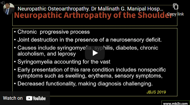
Causes are Diabetes mellitus, Syringomyelia, Spinal cord tumors, Meningomyelocele, Extrinsic compression of the spinal cord, Peripheral nerve tumors, Multiple sclerosis, Congenital insensitivity to pain, Hereditary sensory and motor neuropathy Absenceof normal protective sensory feedback Repetitive mechanical trauma causes progressive joint destruction X rays show hypertrophic joint: manifested radiologically as joint destruction and fragmentation, osseous sclerosis, and osteophyte formation or atrophic…
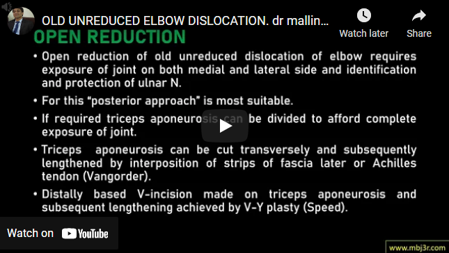
Elbow dislocations are classified according to position of ulna & radius in relation to the distal humerus Post- dislocation account for 80-90% of all dislocations Fall backward on arm with elbow in flexed position with Forearm supinated is most common mechanism Extensive myositis ossificans around joint esp. in brachialis & triceps brachi muscles, Marked shortening of triceps muscle, Shortening of…
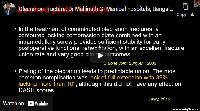
Mechanism of injury : Direct force through the flexed elbow or Hyperextension Olecranon becomes important for elbow stability if the fracture involves more distal part of the anteromedial facet, extending into coronoid process. Investigations : X ray, rarely CT scan required Treatment options include tension band wiring and plate fixation Problem of hardware irritation to skin Complications are Loss of…
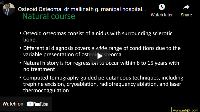
Benign bone tumor (osteogenic) derived from osteoblasts Common sites : Proximal femur (most common),tibial diaphysis, vertebrae Histology – Nidus of osteoid and immature osteoblasts surrounded by a rim of reactive bone formation X- ray shows radiolucent nidus (< 2 cm) surrounded by reactive bone Treatment – observation and NSAIDs, radiofrequency ablation, surgery Osteoid osteoma in spine can cause painful scoliosis, surgical intervention of…
It causes bone to lose strength and density 1 in 2 women over 50 years of age are likely to sustain a fracture due to osteoporosis Osteoporosis accounts for more days in hospitalization than other diseases like DM, MI or breast cancer Spine or hip fractures, are the most serious complications of osteoporosis It is called as silent disease as…