Author page: hanumantha@gravitysoft.in

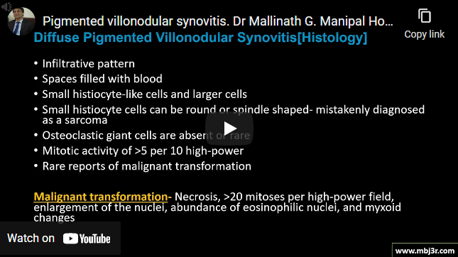
Pigmented villonodular synovitis

Pigmented villonodular synovitis

Benign proliferative disease of the synovial joint, synovial bursa, and tendon sheath Knee joint being involved most often, followed by the hip, ankle, shoulder, and elbow Localized type and  diffuse type Histologic findings of mononuclear cells, giant cells, foam cells and hemosiderin deposits  Histologically may resemble haemophilic joint or GCT of tendon sheath pathognomonic appearance on MRI Arthroscopic or open…
Learn More

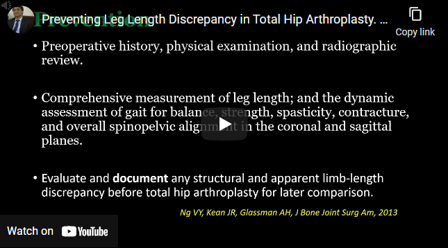
Preventing Leg-Length Discrepancy in THR

Preventing Leg-Length Discrepancy in THR

Limb discrepancy causes patient dissatisfaction, pain, gait disturbance etc Postoperative goal is a final LLD of <1 cm Evaluate and document any structural and apparent limb-length discrepancy before total hip arthroplasty for later comparison Measurement of LLD on a preoperative anteroposterior pelvic radiograph is best performed using the inter-teardrop line and the femoral head center as the landmarks Hip position…

Learn More

Preventing Post-THR Hip Dislocation

Preventing Post-THR Hip Dislocation

Dislocation is the cause of 15% of THA revision surgeries Surgeon factors : Component Positioning, Implant Choice, Surgical Approach Low-volume surgeons and patients with elevated BMI increase the risk of malpositioning No convincing evidence that fluoroscopic or robotic assistance helps  Increasing the size of the femoral head (up to 38 mm) provides greater resistance to dislocation Dual-mobility designs also raise…

Learn More

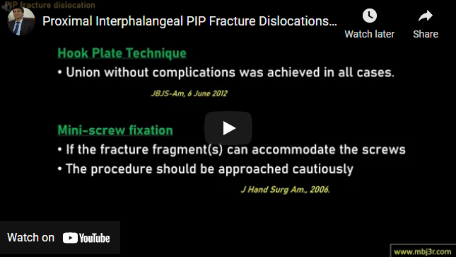
Proximal Interphalangeal (PIP) Fracture-Dislocations

Proximal Interphalangeal (PIP) Fracture-Dislocations

Most common – dorsal dislocation of the PIP joint, associated fracture of the volar articular surface of the middle phalanx.  Most stable when reduced in a flexed position. Dislocations with fractures of <30% of the joint surface that prove stable after closed reduction are buddy-taped to an adjacent finger Extension block pinning is indicated for treating unstable dorsal PIP fracture-dislocation…

Learn More

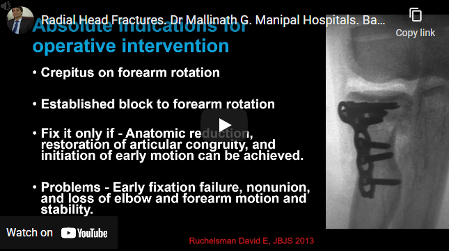
Radial Head Fractures

Radial Head Fractures

The radial head is an important stabilizer for valgus, axial, and posterolateral rotational forces Mason classification Conservative management for Isolated stable Mason type-1 or type-2 fracture of the radial head or neck Indications for operative management are crepitus on movement bone block Surgical exposure: kocher’s approach Low profile plate and headless screws used Protect PIN  Radial head excision typically not…

Learn More

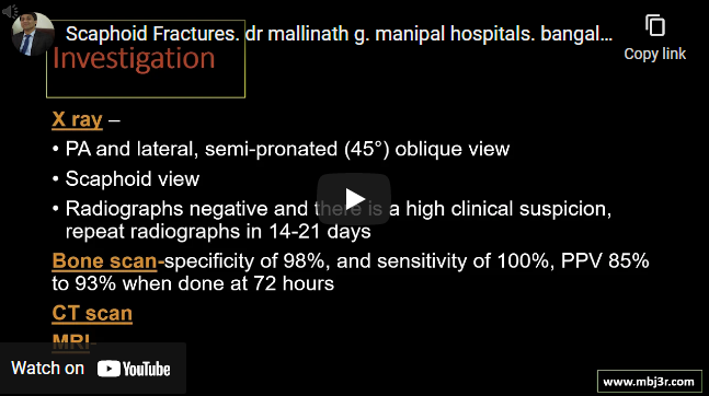
Scaphoid Fractures

Scaphoid Fractures

Dorsal carpal branch of radial artery is major source of blood supply 15% of acute wrist injuries, 60% of all carpal fracture Herbert and Fisher Classification Problematic fracture due to risk of AVN and Non union X ray to be repeated after 2 weeks in suspected cases, till then apply cast MRI and CT to be considered if x rays are…

Learn More