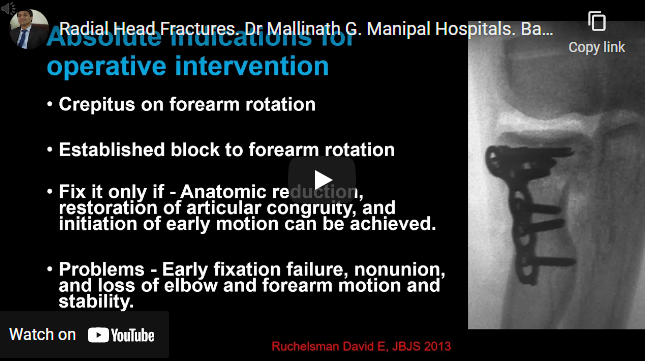
The radial head is an important stabilizer for valgus, axial, and posterolateral rotational forces Mason classification Conservative management for Isolated stable Mason type-1 or type-2 fracture of the radial head or neck Indications for operative management are crepitus on movement bone block Surgical exposure: kocher’s approach Low profile plate and headless screws used Protect PIN Radial head excision typically not…
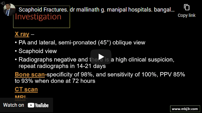
Dorsal carpal branch of radial artery is major source of blood supply 15% of acute wrist injuries, 60% of all carpal fracture Herbert and Fisher Classification Problematic fracture due to risk of AVN and Non union X ray to be repeated after 2 weeks in suspected cases, till then apply cast MRI and CT to be considered if x rays are…
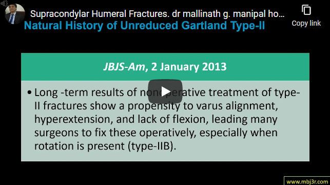
Most commonly seen in children aged 5-7 years with fall on out stretched hand Extension type is seen >95% Gartland Classificaiton is used to classify extension type of SCH fractures Always check for distal pulses If palpable pulses do not return, but the limb is warm and pink and/or pulses are Dopplerable, the patient may be observed for 24 to…
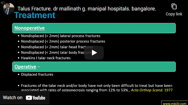
Talar neck fractures most common Mechanism of injury : forced dorsiflexion with axial load Investigations : x-ray and CT scan if needed Non operative management for un-displaced (<2mm) fractures Hawkins classification is used for talar neck fractures Displaced talar neck and/or body fractures should be treated with open reduction and internal fixation Common complications include osteonecrosis, ankle stiffness, arthritis
Moderate to high-energy trauma 8% to 43% of these are open fractures and Ligament disruption seen in 43% to 80% CT scan is done to assess the articular collapse and fracture comminution Schatzker classification is commonly used Treatment is often aimed at restoring the articular congruity, coronal alignment and angular stability Open reduction and internal fixation is the traditional means…