https://www.youtube.com/watch?v=qq9viEFod4k
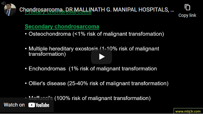
Most common locations include the pelvis, proximal femur, distal femur, scapula Primary vs secondary chondrosarcoma Histologic grade correlates with survival Poor prognostic variables-axial and proximal extremity lesions, advanced patient age, inadequate surgical margins Investigations : MRI,CT and needle biopsy Wide surgical excision is the treatment of choice
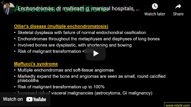
Benign chondrogenic tumor composed of hyaline cartilage caused by an abnormality of chondroblast function in the physis Most common in 20-50 year olds Most common locations hand (60%) > feet Associated conditions: Ollier’s disease (multiple enchondromatosis) and Maffucci’s syndrome Presents with pathological fracture or angular deformities X rays show Pop-corn” stippling, arcs, whorls, rings Symptomatic and Pathologic fractures – immediate curettage and grafting is now favoured Watch out for risk of malignant transformation in…
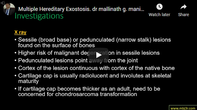
Benign chondrogenic lesion derived from aberrant cartilage from the perichondral ring Solitary and multiple hereditary forms Autosomal dominant inheritance Mechanical symptoms or symptoms of neurovascular compression X Rays show pedunculated lesions point away from the joint cartilage cap becomes thicker as an adult, need to be concerned for chondrosarcoma transformation Symptomatic cases- excision Complications : Vascular compression, Nerve compression, Tendon compression, Chondrosarcoma, Bursa formation
Benign bone tumor (osteogenic) derived from osteoblasts Common sites : Proximal femur (most common),tibial diaphysis, vertebrae Histology – Nidus of osteoid and immature osteoblasts surrounded by a rim of reactive bone formation X- ray shows radiolucent nidus (< 2 cm) surrounded by reactive bone Treatment – observation and NSAIDs, radiofrequency ablation, surgery Osteoid osteoma in spine can cause painful scoliosis, surgical intervention of…
Combination of fractured bone with an existing prosthesis makes these fractures challenging Mortality rate within one year following surgical treatment of periprosthetic femoral fractures is high If the Stem/component loose then revision of the component, with adjunctive stabilization of the fracture with a plate or allograft/plate composite is required. If the Stem/component well fixed then the component should be retained…